
 Community
Tiny Pictures
Ultrasounds and Diagnostic Images During Pregnancy
| Share: | Tweet | Thank You! |
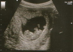 Many women anxiously await the chance to see their baby's tiny fingers and toes on an ultrasound.
Thanks to advanced technology what used to be a simple black and white image of bones and other
difficult to distinguish body parts has turned into a family event for a glimpse of the newest addition.
While it is fun to have an early glimpse at your baby, many women wonder if ultrasounds are safe, and
perhaps even a few more wonder if they even need one at all.
Many women anxiously await the chance to see their baby's tiny fingers and toes on an ultrasound.
Thanks to advanced technology what used to be a simple black and white image of bones and other
difficult to distinguish body parts has turned into a family event for a glimpse of the newest addition.
While it is fun to have an early glimpse at your baby, many women wonder if ultrasounds are safe, and
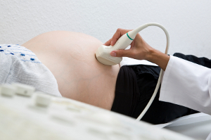
perhaps even a few more wonder if they even need one at all.The truth is that ultrasounds (as is any medical procedure) are optional. Ultrasounds use high-frequency sound waves that are undetectable to the human ear to send waves through the body by the use of a transducer which bounces waves off the baby and then transforms this signal into video and images. While it sounds very high tech, the process is fairly simple and has been in use for many years.
Research has shown that ultrasounds hold no danger to your developing baby no matter how many you have, or how early in the pregnancy. In fact, some high risk pregnancies (those with multiple babies, genetic or birth defects, and others) may have many, many ultrasounds in preparation for a safe birth. Some ultrasounds are conducted over the belly using a conduction gel to help the sound waves pass into the body.
Other women may need a transvaginal ultrasound early in their pregnancy. For women who are around or less than 10-12 weeks, your doctor may use a long probe that is covered in gel which is inserted through the vagina and then used to see the uterus, ovaries and baby. This method is often used for women who are early in their pregnancy to verify that your baby is in fact living and give an estimated due date. Because the uterus does not begin to leave the pelvic bones until around the 10th to 12th week, transvaginal ultrasounds are the only way to get a picture of your growing baby. Transvaginal ultrasounds are painless, and your doctor may ask you to come for your first one with a full bladder to help move your uterus into a better position for pictures.
Ultrasounds are usually performed at two different times during a pregnancy - early, and again at around 20 weeks. If your doctor only does one, that's fine as well... it really just depends on your specific circumstances and the preference of the doctor. There is no right or wrong way to complete them.
An early (or what is sometimes known as a diagnostic ultrasound) is completed usually through the vagina and can help identify:
- More than one baby
- An estimated age of your baby, and the flutter of a heartbeat if you are far enough along (you can expect this on or after the 6th week)
- Diagnose complications or miscarriages
 Your 20 week ultrasound is much more in-depth. This ultrasound is "the big one" that many women
look forward to. If you are interested in finding out if you are having a boy or girl, this is the time to ask.
If you don't want to know, make sure to tell your ultra-sonographer. Your ultrasound will not usually
be conducted by your doctor - typically, a specially trained medical professional known as an ultra-
sonographer will complete the procedure and share a copy of the results with your physician as needed.
Highly trained to spot even a tiny sign of trouble, ultra sonographers are going to be assessing your baby
from head to toe, making sure that every structure essential to healthy growth and development is
present.
Your 20 week ultrasound is much more in-depth. This ultrasound is "the big one" that many women
look forward to. If you are interested in finding out if you are having a boy or girl, this is the time to ask.
If you don't want to know, make sure to tell your ultra-sonographer. Your ultrasound will not usually
be conducted by your doctor - typically, a specially trained medical professional known as an ultra-
sonographer will complete the procedure and share a copy of the results with your physician as needed.
Highly trained to spot even a tiny sign of trouble, ultra sonographers are going to be assessing your baby
from head to toe, making sure that every structure essential to healthy growth and development is
present.During your 20 week ultrasound, you can expect an examination of:
- The placenta (for size and position)
- Baby's position
- Amount of amniotic fluid present
- Gender
- Baby's head size, and estimated length
- A more final estimated due date (based on baby's development thus far)
- Assessment of the heart and all four chambers for defects
- Assessment of abdominal organs (liver, intestines, kidneys, and stomach) for defects
- A look at the spine and brain for size and completeness
Ultrasounds can tell you and your doctor a lot about what has been and what is to come. In the years prior to ultrasound babies were born with conditions that should have never passed through the birth canal. Had there been the opportunity to know and understand what was to come, the medical team could have been more prepared for a safe delivery in whatever method was necessary and there could have been a better outcome for babies and their parents. Remember those stories of "I didn't know I was having twins until the second one was coming out..." That's just one reason ultrasounds can make child birth a more safe process.
Can you choose to not have an ultrasound? Yes, you can. Many women forget that everything is optional in medicine. Should you skip your ultrasounds? No. But the choice is ultimately yours as long as you understand and are willing to accept the risks of foregoing the procedure. Especially given the safety, convenience and benefit of having one, there seems to be no reason not to. If you decline, your physician may ask you to sign a waiver releasing him or her from any liability related to injuries during birth that could have been detected on ultrasound.
No matter if you decide to have your ultrasound or not, childbirth is an exciting time for you and your family. Enjoy every moment, every kick and every nudge until the day you meet face to face.
| Share: | Tweet | Thank You! |
Read & Learn More:
 The Needle's Edge - A Breakdown of Prenatal Testing |
 Sober Mama: Having a Drug-Free Delivery |
 When To Call The Doctor - Things You Shouldn't Ignore During Your Pregnancy |


